UBB strategic research infrastructure elements (rUBB)
Molecular Biology, Biochemistry and Biophysics
Head of laboratory: Prof. Dr. Habil. Horia L. Banciu
UBB Strategic Infrastructure
- Stereomicroscope Axio Zoom.V16 with Fluorescence and Apotome (Carl Zeiss, Germany)
- Guava® Muse® flow microcytometry system for cell culture characterization (Cytek Bioscience)
- Axio Vert.A1 FL inverted microscope with LED fluorescence source and AxioCam 208c color digital camera (Carl Zeiss, Germany)
- FLUOstar Omega Filter-based multi-mode microplate ELISA reader (BMG Labtech, Germany)
- ChemiDocTM Imaging System (Bio-Rad, USA)
- Optical microscope model Axio Scope A1 with fluorescence module and color Axio Cam 105 camera (Carl Zeiss, Germany)
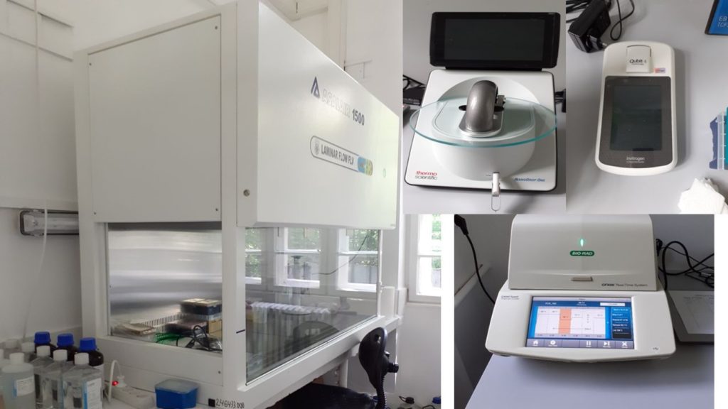
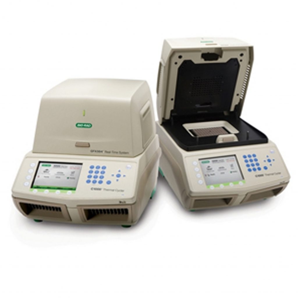
- CFX96 Touch™ Real-Time PCR Detection System (Bio-Rad, USA)
- Server System Intel R1304WFTYS WOLF PASS (Intel, USA)
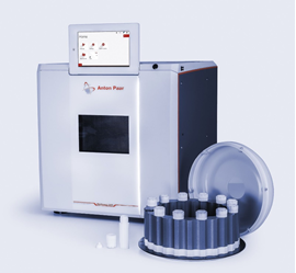
- Microwave digestion system Multiwave 5000 (Anton Paar, Austria)
Stereomicroscope Axio Zoom.V16 with Fluorescence and Apotome (Carl Zeiss, Germany)
Specifications: The AxioZoom V16 is an advanced system with motorized optics for examination of large samples in transmitted, incident and fluorescence light. It allows optical paths to be combined into one, providing images with doubled resolution. The system has motorized focus and zoom functions with a minimum magnification ratio of 16:1, and uses a 120W fluorescent fiber optic source for high quality images. Equipped with two cameras, Axiocam 807 mono and Axiocam 208 color, it provides flexibility in data capture. It includes a structured illumination optical sectioning module, Apotom3, which increases contrast and resolution and eliminates blurred areas. The system comes with image acquisition and processing software, facilitating sequential captures on multiple fluorescence and transmitted light channels. Uniqueness: AxioZoom V16 high-resolution motorized stereomicroscope for transmitted, incident and fluorescent light examination, with digital color camera model Axiocam 208 and monochrome camera model Axiocam 807 mono, with optical sectioning system model Apotome3 and software for analysis of acquired images. Applicability: The AxioZoom V16 is a device used in various scientific fields for microscopic examination of samples. Thanks to its fluorescent light source and the ability to create optical sections by structured illumination, it is ideal for fluorescence microscopy and cell and molecular biology studies. In neuroscience, it allows detailed analysis of neuronal and synaptic structures, facilitating research into brain function and pathology. It is also used in botany, zoology and the pharmaceutical industry, where cells and the effects of drugs are analyzed. AxioZoom V16 is also applied in nanotechnology to analyze microstructures and in pathology to examine diseased tissues. In embryology, it helps to observe the development of embryos and model organisms. Its motorized focus and zoom functions, together with the ability to examine samples in transmitted, incident and fluorescent light, provide high-resolution images, making it an essential tool in research and diagnostics. Fields: Biology, Ecology, Molecular Biology, Molecular Diagnostics, Immunology, Microbiology, Biochemistry, Medical Biology, Geology Teaching provided: Medical Hematology (LR), Hematology and Blood Transfusion (LM), Histopathology (LR). Undergraduate specializations: Biochemistry, Biology and Industrial Biotechnologies; Master: Molecular Biotechnology and Medical Biology (LR and LM), PhD – Doctoral School of Integrative Biology Person in-charge: Access: Monday- Friday, 8-20, by appointment by e-mai

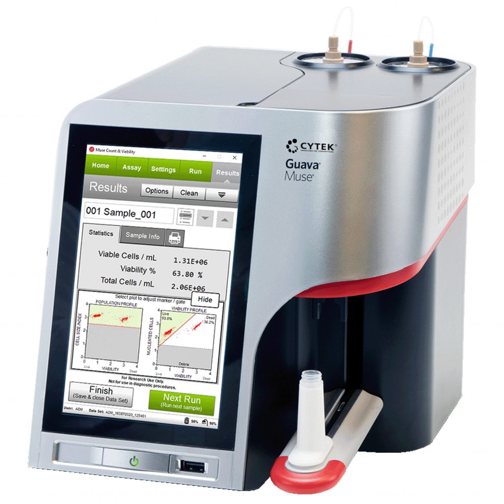
Guava® Muse® flow microcytometry system for cell culture characterization (Cytek Bioscience)
Uniqueness: The Muse® Cell Analyzer is a unique instrument that enables multidimensional cell health analysis on a single platform. It offers a simplified format enables researchers of varying experience levels to obtain a comprehensive picture of cellular health; effortless acquisition and analysis of samples using mix-and-read assays; highly simplified and intuitive touchscreen interface delivers rapid measurements of cell concentration, viability, apoptotic status, and cell cycle distribution, highly sensitive, accurate and rapid detection using minimal cell numbers, superior precision compared to traditional methods. Applicability: cell counting, viability assays, proliferation, apoptosis and oxidative stress, determination of cell cycle phases, study of extracellular and intracellular expression of fluorescently labeled receptors Fields: molecular biology, molecular diagnosis, immunology, microbiology, biochemistry, medical biology Teaching provided: Metabolic biochemistry, Bionanotechnologies; Biochemistry of proteins and introduction to proteomics, Biochemistry of nucleic acids and introduction to genomics; Immunobiology, Medical Biochemistry, Modern Biochemical and Biophysical Methods, (3) Immunobiology; Environmental molecular biology, at the Bachelor’s degree specializations (Biochemistry, Biology and Industrial Biotechnologies) and Master (Molecular Biotechnology and Medical Biology (LR and LM); PhD – Doctoral School of Integrative Biology) specializations. Person in-charge: Access: Mo to Fri, 8-18; with e-mail programming.
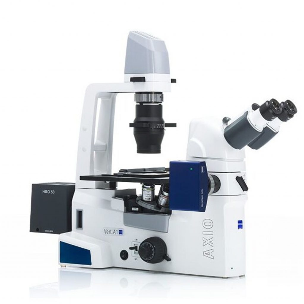
Axio Vert.A1 FL inverted microscope with LED fluorescence source and AxioCam 208c color digital camera (Carl Zeiss, Germany)
Specifications: Axio Vert.A1 FL inverted microscope (Carl Zeiss Instruments, Germany) for examination in transmitted light, light field and phase contrast. State-of-the-art LED fluorescence source. AxioCam 208c color digital camera. Image acquisition and analysis software. Teaching provided: Immunobiology, Modern biochemical and biophysical methods, Bionanotechnologies (Master, Molecular biotechnologies); Environmental molecular biology (Doctoral School of Integrative Biology). Persons in-charge: Access: Mo to Fri, 8-17; with e-mail programming addressed to one of the specialists listed above.
Uniqueness: The inverted microscope with state-of-the-art fluorescence source – Colibri 5, has 4-channel LED for viewing the following fluorochromes: Red: at 630nm, for Cy5 excitations, Alexa 631 nm, Toto-3 and similar colors; Green: at 475 nm, for Cy3, TRITC, DsRED excitations and similar colors; Blue: at 475nm for eGFP, Fluo4, FITC excitations and similar colors; Ultraviolet: at 385 nm for DAPI, Alexa 405, Hoechst 33258 and similar colors. It has the option of generating high resolution images with overlapping colors. The microscope has ECO Power function, a 8.3 Megapixel AxioCam 208c digital camera and image acquisition and analysis software (ZEN 3.0 lite + ZEN lite / starter Module Measurement).
Applicability: cell counting, observation of mammalian cell morphology in 2D and 3D culture, intracellular uptake studies for different fluorescent compounds, cell apoptosis studies, medical diagnostic methods (immunofluorescence), examination of biological fluids or other tissues (from plants, animals, human).
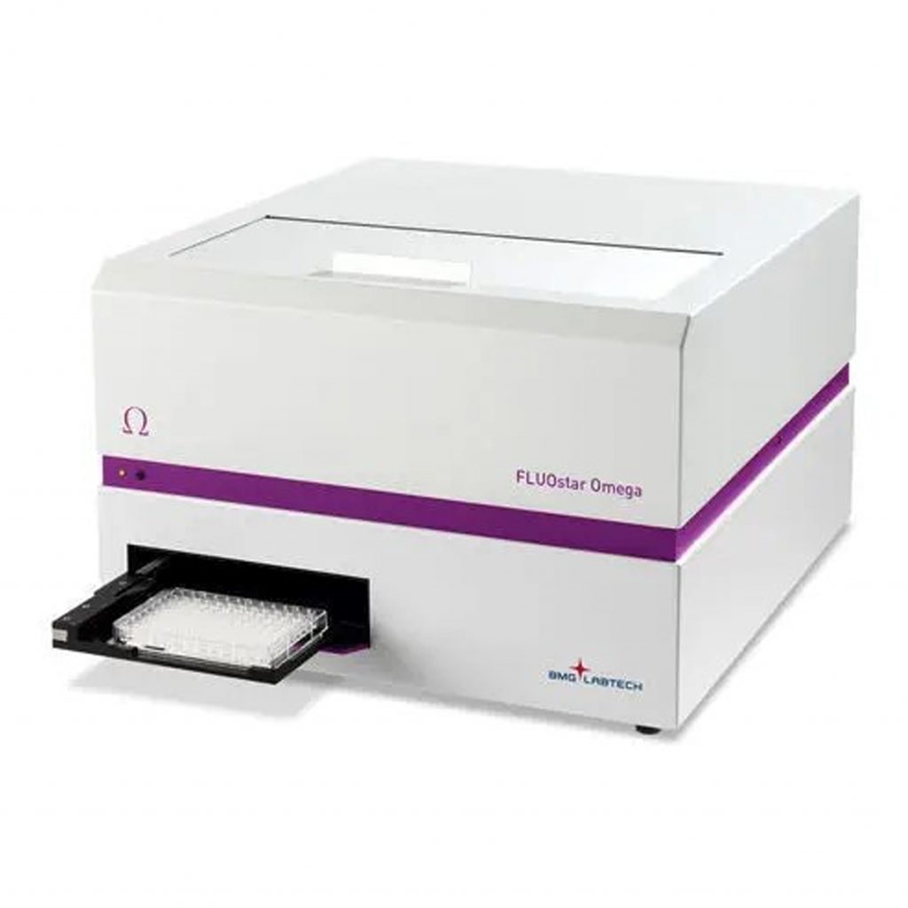
FLUOstar Omega Filter-based multi-mode microplate ELISA reader (BMG Labtech, Germany)
Specifications: The FLUOstar Omega multi-detection microplate reader provides the following detection modes: Ultra-fast UV/vis absorbance spectra or filter-based absorbance; fluorescence intensity (allows the use of multi-excitation and multi-emission applications, such as FRET, BRET, FURA-2 and other ratiometric methods); luminescence (flash & glow). Uniqueness: The FLUOstar® Omega is is built upon BMG LABTECH’s unique Tandem Technology. This is a combination of two technological concepts – an ultra-fast, full spectrum absorbance spectrometer, and extremely sensitive filter-based detection, with advanced optics and a photomultiplier tube to provide superior sensitivity for all detection modes. Applicability: detection and quantitation of antibodies against clinically relevant molecular markers, quatitation of endotoxins, nucleic acids, proteins, measurement of enzyme activity, ATP quantitation, microbial and animal cell growth monitoring by optical density. Fields: molecular biology, biochemistry, molecular diagnostics, immunology, microbiology. Teaching provided: Metabolic Biochemistry, Immunobiology, Biochemistry of Proteins and Introduction to Proteomics, Biochemistry of Lipids and Carbohydrates (undergraduate – Biology, Biochemistry); Modern Biochemical and Biophysical Methods, Bionanotechnology (Master – Molecular Biotechnology), Medical Biochemistry, Molecular Immunology (Master – Medical Biology) Access: Mo to Fri, 8-17; with e-mail programming addressed to one of the specialists listed above.
Persons in-charge:
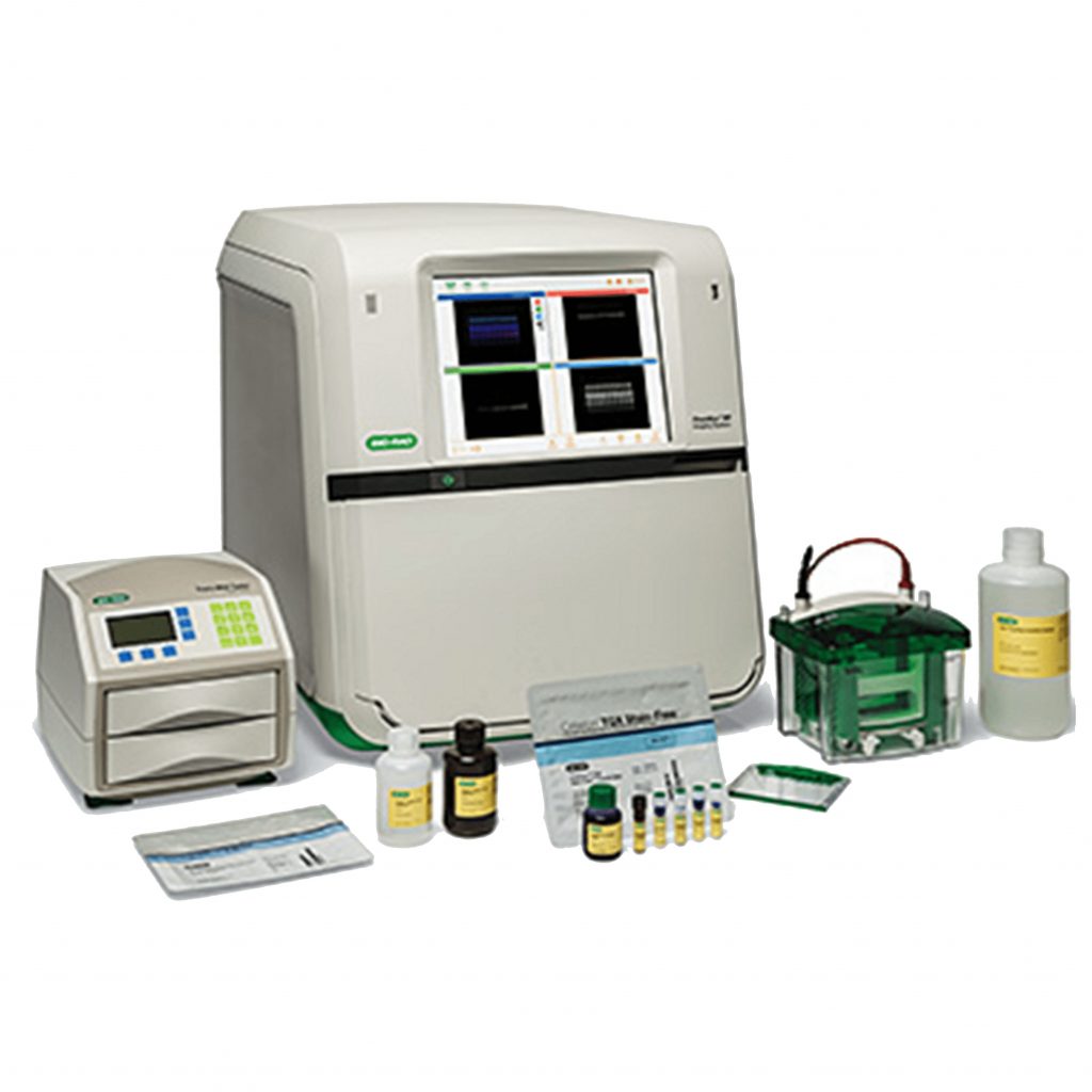
ChemiDocTM Imaging System (Bio-Rad, USA)
Specifications: The ChemiDoc Imaging System provides fast, reliable, and sensitive imaging and documentation of gels and chemiluminescence Western blots. This system is compatible with stain-free technology, chemiluminescence detection, and a wide range of gel stains such as ethidium bromide, SYPRO Ruby, Coomassie, and silver stains. The ChemiDoc Imaging System can be upgraded to gain the full multiplex fluorescent Western blotting capabilities
Applicability: nucleic acid and protein research.
Fields: molecular biology, proteomics, immunology, genetics, molecular diagnosis.
Teaching provided: Metabolic biochemistry; Biochemistry of proteins; Biochemistry of nucleic acids, Immunobiology (undergraduate – Biochemistry); Recombinant DNA Technologies, Medical Biochemistry (Master – Molecular Biotechnology and Medical Biology); Environmental Molecular Biology (PhD – Doctoral School of Integrative Biology).
Person in-charge: Lecturer Dr. Alina Sesărman – alina.sesarman[a]ubbcluj.ro
Access: Mo to Fri, 8-17; with e-mail programming.
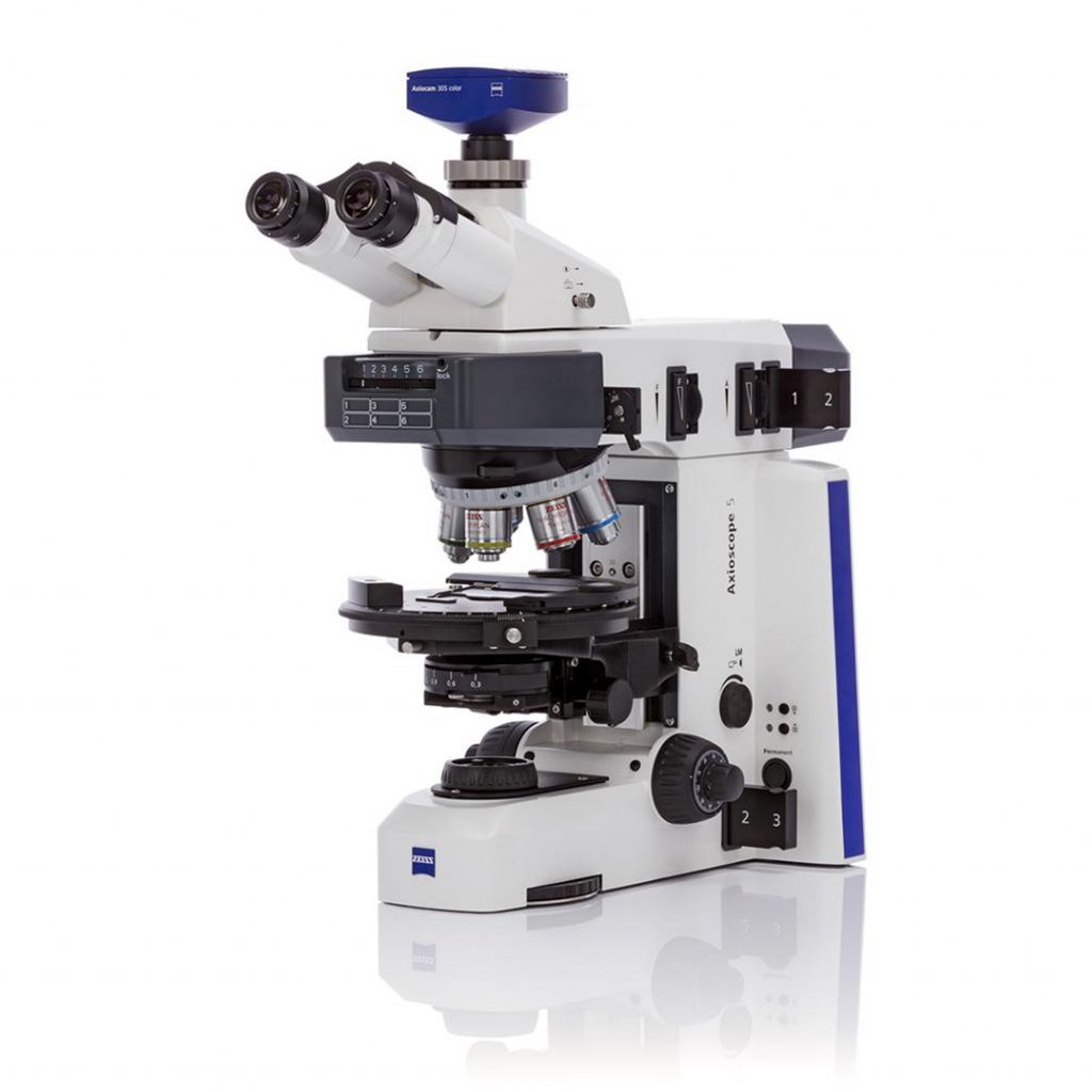
Optical microscope model Axio Scope A1 with fluorescence module and color Axio Cam 105 camera (Carl Zeiss, Germany)
Specifications: Axio Scope A1 Microscope HAL 50, FL-LED, 3x H Microscope, 3x DIC with Fluorescence Module (365 nm and 540 nm fluorescence), Axio Cam 105 color camera with CMOS sensor, 5 Mpixels resolution
Uniqueness: Optical microscope with multiple functions (direct light, phase contrast, dark field, fluorescence)
Applicability: cell count, cell morphology, cellular structures (fungal, animal and plant cells), cell viability assessment.
Fields: environmental and medical microbiology, cell biology, animal cell cultures, hematology, animal / plant histology, histopathology.
Teaching provided: Biophysics (undergraduate – Biology, Biochemistry); Modern Biochemical and Biophysical Methods (Master – Molecular Biotechnology)
Persons in-charge:
- Prof. Horia Banciu – horia.banciu[a]ubbcluj.ro
- Lecturer Dr. Emilia Licărete – emilia.licarete[a]ubbcluj.ro
- Teaching Assist. Laura Pătraș – patras.laura88[a]yahoo.com
Access: Mo to Fri, 8-17; with e-mail programming addressed to one of the specialists listed above.
CFX96 Touch™ Real-Time PCR Detection System (Bio-Rad, USA)
Specifications: C1000 Touch ™ thermocycler model with CFX96 ™ optical module (Bio-Rad, Hercules, CA, USA). Optical detection: Excitation 6 filtered LEDs; Detection of 6 filtered photodiodes; Range of excitation / emission wavelengths 450-730 nm; Detection sensitivity 1 copy of the target genomic DNA sequence; dedicated software for data analysis.
Applicability: amplification and quantification of gene sequences, melting curve analysis, gene expression analysis, allelic discrimination.
Fields: molecular biology, genetics, molecular diagnosis, genomics, transcriptomics.
Teaching provided: Biochemistry of nucleic acids with genomic elements (undergraduate – Biochemistry); Structure and evolution of the genome (Master – Molecular Biotechnology); Environmental Molecular Biology (PhD – Doctoral School of Integrative Biology).
Persons in-charge:
- Prof. Horia Banciu – horia.banciu[a]ubbcluj.ro
- Lecturer Dr. Alina Sesărman – alina.sesarman[a]ubbcluj.ro
Access: Mo to Fri, 8-17; with e-mail programming addressed to one of the specialists listed above.
Server System Intel R1304WFTYS WOLF PASS (Intel, USA)
Specifications: Processor Xeon® Gold 6252 24C/48T 150W 2.10G 35.75M 10.40GT/sec ITT, Memory 12 X 64GB_LRDIMM_2666
Applicability: NGS data analysis, genome assembly, phylogenomics, comparative genomics, metabolic reconstruction
Fields: advanced bioinformatics, molecular biology, molecular ecology, biodiversity, medical genomics and proteomics, interactomics.
Teaching provided: Biochemistry of nucleic acids with genomic elements & Introduction to Bioinformatics (undergraduate – Biochemistry); Structure and evolution of the genome & Bioinformatics (Master – Molecular Biotechnology); Environmental Molecular Biology (PhD – Doctoral School of Integrative Biology).
Persons in-charge:
- Prof. Horia Banciu – horia.banciu[a]ubbcluj.ro
Access: Mo to Fri, 8-17; with e-mail programming addressed to one of the specialists listed above. Remote access of the server is possible upon providing the appropriate username and password.
Microwave digestion system Multiwave 5000 (Anton Paar, Austria)
Specifications: The equipment allows simultaneous mineralization of 12 samples (rotor 12HVT) of soil, water, organic substances (plants, fungi, bacteria, food, feed), materials, petrochemical samples, plastics, polymeric materials, cosmetics, pharmaceuticals, metals, alloys and geochemicals for evaluating the content of nutrients, substances in excess, toxic. It allows analysis starting from 0.2 g, which is important for samples for which the quantity is limited (e.g. toxicity experiments). Ambient temperature range up to 180 oC.
Uniqueness: SMART VEN ventilation technology, with sensor system that allows automatic detection of each reaction vessel, as well as rotor revolution monitoring, uniform microwave heating and prevention of localized overheating; IR sensor temperature control that provides different AVG, MAX, MIN temperature control strategies. Internal Temperature control in all positions / SmartTemp. All this ensures the efficiency of mineralization and increased safety for the personnel using the device. The short temperature rise and fall time ensures the rapidity of the mineralization process and allows the device to be used for more samples/day than in the classic sand bath mineralization system. Built-in standardized methods over 500 methods, simplify the flow. Directed Multimode Cavity for the shortest heating times in a small-footprint system. TURBO heating and cooling for the shortest overall process times. One-vessel digestion mode for low-throughput applications. Extremely lightweight aluminium rotor (5 kg): as an optimized integral part of the Directed Multimode Cavity, no deformation, no corrosion, no loss of stability.
Applicability: mineralization of soil samples, water and organic substances (plants, fungi, bacteria, food, feed), testing of materials, as well as petrochemical, plastic, polymer, cosmetic, metal, alloy and geochemical samples for the assessment of nutrient content, excess or toxic substances.
Fields: Geochemistry, Environment, Plant Biology, Environmental Biology, Environmental and Medical Microbiology, Cell Biology, Ecotoxicology, Physiology.
Teaching provided: Plant Biochemistry and Molecular Biology, Ecological Biochemistry, Ecophysiology (Bachelor – Industrial Biotechnologies, Biology, Biochemistry); Plant Molecular Biochemistry and Physiology (Master – Molecular Biotechnology), Environmental Molecular Biology (PhD – Doctoral School of Integrative Biology).
Persons in-charge:
· Conf. dr. Dorina Podar – dorina.podar@ubbcluj.ro
· Drd. Lorena C. Văcar – cristina.vacar@ubbcluj.ro
· Drd. Emanuela Dana Tiodar – emanuela.tiodar@ubbcluj.ro
Access: Mo to Fri, 8-16; with e-mail programming addressed to one of the specialists listed above.
Other infrastructure of the laboratory

